Tuesday, July 31, 2018

Natto

Why the Japanese Love NATTO and You Should Too

by John Moody Affiliate linksHealthy LivingSacred Foods
natto benefits
The world of soy can be very confusing for consumers! Most soy based foods are downright dangerous especially for babies, children and those with thyroid disease. 
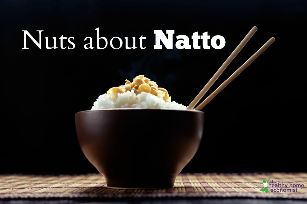
This would include modern concoctions such as soy formulasoya milkbean curd or tofu along with the wide variety of processed foods containing this legume. It is a very important additive for the food industry because it is cheap to produce, replacing more expensive ingredients!
Dozens of scientific studies confirm the dangers of soy in the diet, although the soy industry has predictably countered with “science” of its own featuring cherry picked data and skewed statistics.
While most soy foods are deserving of a warning label, a few others are actually healthy such astraditional soy sauce, miso, tempeh and natto. These four lone exceptions have a long history of traditional use in various Asian diets. They also have good research supporting a positive role in human health.
Of these, the healthiest by far is natto. It is also the least popular, for reasons discussed further below.

What is Natto?

What do these few soy foods have in common? Generally speaking, it is fermentation. Soy is full of a host of anti-nutrients – phytates, goitrogens, phytoestrogens and more.
The truth is that ancient people groups didn’t consume much soy until they discovered the power of fermentation to unlock the nutritional potential by reducing the large amount of anti-nutrients that persist even after cooking for long periods of time.
This discovery occurred around a thousand years ago in China. Someone took some boiled soybeans and added an unknown colony of beneficial bacteria to it. About 24 hours later, natto was born!
Most likely, this happened by either accidental or purposeful contamination of the cooked soybeans with stray bacterial flora in the surrounding environment. A millennium ago, straw bags often served as the equivalent of lunch sacks in some places! This type of container would have contained probiotics that occur naturally on the surface of all living things.
A similar occurrence in the folklore apparently happened with the discovery of kefir. Shepherds in the Caucasus mountains observed that fresh milk carried in leather pouches would ferment into the tangy, effervescent beverage. Once again, the magical process took about 24 hours.

Why Fermented Soybeans are a Superfood

Today, natto is primarily enjoyed by the Japanese, who commonly enjoy a small dish of fermented soybeans with breakfast.
Modern science has shed light on natto’s nature and how it plays a powerful role in bolstering the health of this long lived culture. The bacteria Bacillus subtilis is responsible for turning the cooked soybeans into a superfood. This strain is also known by the common names hay bacillus or grass bacillus.
People have to lend a hand along the way, though. First, they must either boil or stream the beans. Then, they must inoculate them with the beneficial bacteria.
Last, the mixture needs time to ferment. Usually about 24 hours – a bit longer if temperatures are cool.
In the early 1900s, researchers figured out how to make the starter culture apart from using straw. This innovation allowed more consistent results in homemade natto production and the first large scale commercial production as well.
Natto is now enjoyed all over the world as a traditional food with a wide array of health benefits for those who consume it regularly.

Health Benefits of Natto

Natto is one of the few plant based food options that is truly nutrient dense. It is high in a number of minerals – manganese, iron, copper, potassium, phosphorus, and zinc. Since it is fermented, it is also an excellent source of vitamin K2, which few whole foods contain. Rampant K2 deficiency exists in the modern diet for this reason with deadly results.
In fact, natto is the highest food in Vitamin K2 on the planet! People who eat it regularly avoid the need for Vitamin K2 supplements.
How much does natto contain? Up to 100 times more K2 than many cheeses, which are the most accessible food in the Western diet that contains significant amounts of this nutrient. Gouda and brie are some of the highest.

Why is Vitamin K2 so Valuable?

Vitamin K2 is the kind of K our bodies desperately need. Similar to how our body needs true vitamin A, and not just beta carotene, K1 may get turned into K2 by our body.
Unfortunately, for genetic or other reasons such as gut imbalance, it frequently doesn’t happen that way. So, while plants are sometimes great sources of Vitamin K1 and many other precursors to nutrients our body needs, it doesn’t mean that we end up with the nutrients we need such as Vitamin K2.
Current estimates are that over 90% of people are deficient in Vitamin K2. Deficiency can manifest in a wide variety of ways such as excessive wrinkling of the skin, bone loss issues, cardiovascular disease, and cancer.
Since bacteria make K2 via fermentation, many fermented foods, especially natto, contain large amounts of it. Meat, dairy, eggs, and other animals foods contain varying amounts of K2. For those who eschew these foods or otherwise lack access to them, natto is one of the very few options to choose from to ensure sufficient K2 in their diet.
Even for those eating animal products, because few eat “nose to tail” organ meats, they may not get enough vitamin K2. So, natto can play a crucial role as a low cost, truly natural whole food that has many other benefits to boot.

How Much Natto to Eat and How Often?

Eating only a few ounces of natto equates to taking 5-6 (expensive) Vitamin K2 supplement capsules! A single 100 gram (3.5 ounce) serving contains 775 micrograms. Science shows that eating natto most definitely improves blood amounts of this crucial vitamin too. (1)
Thus, eating a serving of natto once or twice a week would provide plenty of K2 in the diet.

Natto Probiotics

Beyond its nutritional value, properly prepared natto is a powerful probiotic. Just be sure not to cook it else you destroy this major health benefit!
The final product is bursting with literally billions of beneficial bacteria. Studies have shown that the strains used to make natto may help with irritable bowel and ulcerative colitis among other health issues. (23)
A number of studies show that natto, both its probiotic bacterial component and other compounds the final product contains, promote proper immune function and other immune system benefits. (45)
Some doctors think that, unlike many other probiotics, natto may survive the body’s natural digestive defenses and help colonize our lower bowels with beneficial bacteria.

Nattokinase

As a living food, natto brings one other benefit worth mentioning to the dinner table – enzymes. Health conscious people are familiar with the concept of popping digestive enzyme supplement after a meal of cooked or processed food to help nutrient assimilation and ward off unpleasant symptoms like bloatingand heartburn. Why not just eat food that already contains them for a less expensive and more effective approach?
Natto contains one enzyme in particular, aptly named nattokinase, that shows numerous benefits for the cardiovascular system in particular. The enzymes found in natto also show possible health and other benefits beyond digestive according to Dr. Ralph Holsworth, an emergency room supervisor and biomedical researcher in a rural hospital in Colorado.
Dr. Holsworth has coauthored several studies on the enzyme nattokinase, a byproduct of natto fermentation. He says that this enzyme:
breaks down fibrin in the blood, a protein aggregate involved in blood clotting, decreases the ‘stickiness’ of the red blood cells, and assist in the prevention of arterial plaque formation.
These blood-thinning actions may lessen the severity of heart attacks and other cardiovascular events. (6)
Dr. Holsworth uses nattokinase in his own medical practice to help prevent blood clots and assist in healing from surgery. Nattokinase is not yet widely used in mainstream medical practice, although it has been gaining popularity as a food supplement with the public. (7)
So, with natto you get a food with an excellent nutrient profile plus many other known benefits. Yet, that doesn’t mean natto presents no dangers or problems.

Natto Dangers

While natto is a traditional food, modern soybeans are not your great, great grandma’s soybeans. In fact, soy is one of the worst of the worst of modern, industrialized crops. This means a high level of genetic modification such that you can literally douse the crops with Roundup without them dying!
Not just glyphosate containing herbicides either! Soy is endlessly sprayed with a myriad of other industrial agricultural chemicals. Just ask anyone who lives anywhere near a soybean farm!
After harvest, the soybeans are then processed with more toxic chemicals such as the known carcinogen hexane. (8)
Modern soy especially frankenfoods like seitan and soy protein isolate don’t come close to resembling their ancient roots. They don’t have the same health benefits by a long shot even when consuming it in whole form such as edamame.
Count on it having a great deal of chemical and pesticide residue contamination when it reaches store shelves.

Natto May Contribute to Allergies

One last note, natto (and soy sauce) is high in histamines. This is a chemical group common in fermented foods that are linked to things like allergies, acne and other skin related issues like eczema and rosacea.
They pop up whenever you have an immune response, helping eliminate the offending invader. Thus, histamines are both good and good for you. But some people overproduce histamines or suffer fromhistamine intolerance. This contributes to many issues like seasonal allergies and other autoimmunity problems.
If you are one of these people, you may still be able to eat natto and soy sauce. For some, you may just need to eat it infrequently or in very small amounts. Others may find they can’t tolerate it at all.

Organic Natto

With natto, it is very important to stick with an organic, traditionally made brand. Traditional natto is made only from soaked, then fermented whole soybeans.
Also note, most modern, mass produced natto is sold in styrofoam (polystyrene) packaging. This stuff is an environmental disaster and may put nasty stuff into the food it contains.
Look for organic natto in safer packaging options. Most good quality nattos will come in safer plastic or glass packaging. Asian grocery stores in the refrigerated or freezer section are a good place to look.

Taste and Texture Quite Unlike Any Other Food!

Like many fermented foods, natto is an acquired taste to put it mildly. Some people never get used to it, in fact! It has a very pungent aroma, along with a rather unappealing texture and taste.
These three strikes against it greatly impact what should be a highly nutritious food that most anyone can afford. Some people do seem to enjoy the taste, though, so don’t automatically assume you won’t like it. As one sampler put it, natto was at best, “Klingon food.” (9)
As the saying goes … don’t knock it until you try it!

Best Ways to Go Nutty with Natto

Since natto is a something that probably requires getting used to, if you want to try it, the safest and surest bet is to mix it with rice or soup. Don’t ever cook it though. Be sure to blend in once the mixing food has cooled a bit. Mixing in a dollop of cultured cream with a bowl soup at the table involves a similar process.
This recipe for natto fried rice is a good place to start.
Small amounts of natto (remember: you don’t need a lot to reap huge health benefits!) often does best when paired with a larger proportion of a relatively bland food. This effectively helps to disperse its strong smell and hide the unpleasant texture.
While mass produced natto is now easy to find, there are many local and regional options that are probably far better to support with your food dollars.
Start with a traditionally made, organic variety. Otherwise, you are likely getting GMO soy along with some glyphosate residue and possibly undesirable additives too.
Have you tried natto and lived to tale the tale? Do you eat it regularly and if so how so? What ways do you find to eat this traditional food that overcomes the strong smell and texture?
John Moody is an author, speaker, farmer, homesteader, and Real Food activist. Most importantly, he is husband to an amazing wife and five awesome kids. John speaks nationally at a wide range of events, along with writing for numerous publications and consulting for farmers, homesteaders, and food businesses.

One in Nine Adverse Events Reported After DTaP is seerious

JULY 31, 2018

One in Nine Adverse Events Reported After DTaP Vaccination is Serious—But CDC Says, “Don’t Worry, Be Happy”

By The World Mercury Project Team

Until the 1990s, the vaccine administered to children for diphtheria, tetanus and pertussis protection was the DTP vaccine, one of the first combination vaccines ever licensed by the U.S. Food and Drug Administration (FDA). However, as a “whole-cell” vaccine (meaning that it contained the entire Bordetella pertussisorganism rather than purified components), DTP had a significant downside—including published safety concerns dating back to the 1930s and widespread reports of neurological damage emanating from both the United States and other countries. By 1991, the Institute of Medicine cautiously reported that the evidence was “consistent with a possible causal relation between DTP vaccine and acute encephalopathy” [brain disease].
…characterized pertussis prevention as ‘an unresolved problem,’ noting the ‘progressive increase’ in pertussis incidence after introduction of the acellular vaccines and the need for ever more boosters.
To pacify a concerned public, the Centers for Disease Control and Prevention (CDC) advised a phase-out of the whole-cell vaccine around 1991, while promoting an “acellular” version called DTaP (diphtheria, tetanus and acellular pertussis). By 1997, the switch had taken place for all five doses in the series, recommended for infants and children at two, four, six and 15-18 months and 4-6 years. In the two decades since the changeover, however, the DTaP vaccine has been plagued by embarrassingly low effectiveness. A 2018 article characterized pertussis prevention as “an unresolved problem,” noting the “progressive increase” in pertussis incidence after introduction of the acellular vaccines and the need for ever more boosters. Another recent commentary flatly stated that “pertussis is…not under control in any country” and that new types of pertussis vaccines are needed.
Nonetheless, on the safety front, health authorities have regularly praised the DTaP vaccines as offering a safer alternative than their whole-cell predecessors. Is this reputation for safety well-deserved? CDC researchers writing in June 2018 in Pediatrics seem to think so—but a closer reading of their findings suggests otherwise.

Examining DTaP’s track record

For their study, the CDC researchers assessed over two decades’ worth of data (1991–2016) from the CDC- and FDA-administered passive surveillance system called VAERS (Vaccine Adverse Events Reporting System), examining adverse events (AEs) reported to VAERS following vaccination with one of five currently licensed DTaP vaccines (see table). The five vaccines included two DTaP-only vaccines (approved for the full five-dose series of shots) and three combination vaccines (approved for some portion of the DTaP series). The combination formulations in question included DTaP plus hepatitis B vaccine (HBV), inactivated polio vaccine (IPV) and/or Haemophilus influenzae type b (Hib) vaccine.
The researchers used several methods to consider DTaP vaccination risks, including 1) compiling all “serious” and “non-serious” adverse events reported to VAERS in association with the five vaccines over the designated time period; 2) clinically reviewing all deaths reported to VAERS following DTaP vaccination; 3) reviewing a subset (5%) of “non-death serious reports”; and 4) running an automated search of reported anaphylaxis following DTaP vaccination.

Not so safe

The analysis of VAERS reports identified tens of thousands of AEs (N=50,157) in the aftermath of a DTaP-containing vaccine. (A single VAERS report may include more than one AE, so the adverse event categories are not mutually exclusive.) VAERS, by the federal government’s own admission, captures only about 1% of AEs; thus, the 50,000-plus AEs probably vastly underrepresent the number of real-world DTaP-related vaccine injuries.
The study’s results illustrate the heavy burden of vaccines to which children in the U.S. are subjected. For about 88% of the VAERS reports analyzed, children received the DTaP vaccine concurrently with one or more other vaccines, even though the five types of DTaP vaccine in and of themselves already constitute potent combinations. Researchers who have looked at the number of vaccines administered at well-child visits have pointed out that American infants receivemore vaccines in their first year than infants anywhere in the world.
…many vaccines (including DTaP) are administered in bundles at health care visits around two and four months—exactly when nine out of ten SIDS deaths occur.
Roughly one in nine (11.2%) of the reported AEs were coded as serious, and 15% of all serious AEs were deaths (844/5,627). (If one were to average these deaths over the 26 years from 1991 through 2016, this would represent over 32 deaths annually.) Of note, the investigators’ perusal of death certificates, autopsy reports and medical records showed that the reported cause for nearly half of the deaths (48.3%) was sudden infant death syndrome (SIDS), nearly always in children under six months of age. Although the researchers dismiss the possibility of a causal relationship between vaccination and SIDS, evidence from other corners is strongly suggestive of just such a link. In fact, it strains credulity to deny a plausible connection: many vaccines (including DTaP) are administered in bundles at health care visits around two and four months—exactly when nine out of ten SIDS deaths occur.
Serious but non-fatal AEs cited in 10% to 35% of all VAERS reports included systemic symptoms such as pyrexia (fever), vomiting, seizures/convulsions, diarrhea, lethargy and hypotonia (muscle weakness). Anaphylaxis occurred far less frequently, but most reported anaphylactic reactions arose quickly—within 30 minutes of vaccination. Seizures were the fourth most common serious AE reported. Other studies have detected a heightened risk of febrile seizures when children receive DTaP simultaneously with other vaccines. Febrile seizures are not benign (as once thought), which makes the frequency of post-DTaP seizures concerning.
The authors do not explain why they counted pyrexia as both a serious and nonserious AE, but it accounted for one in five of the latter. As a potential sign of drug allergy and an indicator of a “systemic inflammatory response to a stimulus such as infection,” pyrexia and its prominence are noteworthy. Back in 2004, other CDC researchers commented on the difficulty of ascertaining “the true importance of fever as an [adverse event following immunization]” and noted a lack of clarity regarding “how to interpret fever data derived from vaccine safety trials or immunization safety surveillance.”

What the study leaves out

Although the CDC authors noted that their analysis excluded Quadracel, the most recently approved combination DTaP-IPV vaccine (licensed in 2015), they curiously do not explain why they omitted several other licensed DTaP vaccines that were in widespread use during the time period in question:
  • The Tripedia vaccine (manufactured by Connaught, which through a series of mergers became Aventis Pasteur and later Sanofi Pasteur) was approved as a fourth and fifth DTaP dose in 1992, 1996 and 2000; in 2001, Aventis Pasteur reformulated Tripedia and the FDA approved it for all five doses.
  • Acel-Imune (manufactured by the now-defunct Lederle Laboratories) was approved for the fourth and fifth DTaP doses in 1991 and, in 1996, for the full five-dose series.
  • The Certiva DTaP vaccine (made by North American Vaccine Inc., which was acquired in 2000 by Baxter International Inc.) was licensed in 1998 for doses one through five.
The authors also neglect to mention that all five DTaP vaccines included in their review contain one or more neurotoxic aluminum adjuvants, along withformaldehyde and polysorbate 80, a stabilizer for which information on potential chronic health effects is “not available.” The Tripedia vaccine that the study excluded featured both aluminum and the mercury-containing preservative thimerosal. Adverse events reported during post-approval use of Tripedia included “idiopathic thrombocytopenic purpura, SIDS, anaphylactic reaction, cellulitis, autism, convulsion/grand mal convulsion, encephalopathy, hypotonia, neuropathy, somnolence and apnea.” By excluding these other acellular DTaP vaccines, the CDC study underestimates the magnitude of DTaP-related adverse reactions still further.

Weighing the risks

The CDC authors wrap up their assessment of DTaP vaccine safety with the boilerplate pronouncement that their analysis “did not identify any new or unexpected safety issues.” Parents might disagree, wondering whether it makes sense to expose their child to a not-insignificant risk of serious DTaP-related injury when the risk of diphtheria is virtually non-existent in the U.S. (zero cases in 2016) and the risk of tetanus is likewise minuscule. (Tetanus, in any event, is non-communicable.)
… pertussis incidence has steadily increased (not decreased) in the U.S. since 1980, despite high vaccine coverage.
Evaluating the risks of pertussis infection versus pertussis vaccination in different age groups is somewhat more complex but requires admitting up front that pertussis incidence has steadily increased (not decreased) in the U.S. since 1980, despite high vaccine coverage. Discussing the problem of waning immunity, a 2012 study reported that “after the fifth dose of DTaP, the odds of acquiring pertussis increased by an average of 42% per year.” In fact, the track record for whole-cell and acellular pertussis-containing vaccines shows that both are fraught with problems. Back in 1993, researchers writing in the New England Journal of Medicine observed that a pertussis epidemic in Cincinnati had “occurred primarily among children who had been appropriately immunized” with the whole-cell vaccine. The same pattern of pertussis outbreaks in fully vaccinated populations has occurred with the acellular vaccines. A related but underacknowledged problem is the role of vaccinated individuals as asymptomatic carriers and reservoirs of infection for vulnerable infants. Finally, some researchers have suggested that pertussis vaccination may result “in selection of more virulent strains that are more efficiently transmitted by previously primed hosts.” Specifically, the acellular vaccines only contain B. pertussis antigens “that hold little or no efficacy against B. parapertussis,” which is another causative agent of pertussis infection; researchers concluded in 2010that acellular vaccines “interfere with the optimal clearance of B. parapertussis” and may “create hosts more susceptible to B. parapertussis infection.”
Whether one focuses on safety or effectiveness, it is apparent that simplistic slogans and Pollyanna attitudes are no help in evaluating vaccine risks and benefits. Ultimately, it should be up to parents—not CDC researchers biased against a fair consideration of risks—to make their own informed vaccine decisions.
Mini Review
Volume 2 Issue 3 - 2015
The Unsuspected Capacity of Melanin to Transform Light Energy into Chemical Energy and the Surprising Anoxia Tolerance of Chrysemys Picta
Arturo Solis Herrera*
Centro de Estudios de la Fotosintesis Humana, México
Received: July 19, 2015 | Published: August 24, 2015
*Corresponding author: Arturo Solis Herrera, Centro de Estudios de la Fotosintesis Humana, México, Tel: 014499160048, 014499150042; Email: 
Citation: Herrera AS (2014) The Unsuspected Capacity of Melanin to Transform Light Energy into Chemical Energy and the Surprising Anoxia Tolerance of Chrysemys Picta. MOJ Cell Sci Report 2(3): 00031. DOI:10.15406/mojcsr.2015.02.00031

Abstract

Oxygen is considered critical to nearly all life on earth, as the end electron acceptor that makes, theoretically, mitochondrial oxidative phosphorylation possible, and thereby energy production. Anaerobic energy sources can only temporarily supply ATP and maintain cellular function before substrate depletion, energy shortfall, or end-product poisoning that threaten survival. In most vertebrates, the limits of anoxia tolerance are short, of the order of minutes, because of the urgent dependence (theoretically) of the heart and central nervous system on a continuous supply of O2.
Because greater than 50% of the energy consuming processes of the normoxic cell, especially neuron can be accounted for by ion pumping, a decrease in membrane ion leakage can contribute in a major way to energy savings. Theoretically, the power to the inside of the cell depends on ATP, but in the light of our discovery of the intrinsic property of melanin transform light energy into chemical energy by means of the water molecule dissociation, we think that the levels of ATP, in turn; depend almost completely from the energy that emanates from the melanin.
The purpose of discerning the ability of the painted turtle, of tolerating extended periods of anoxia, even months, will improve our understanding of the pathophysiology and progress in therapeutic strategies in diseases that can be classified as events hypoxic severely affecting human beings, such as cerebral vascular events.
Keywords: Melanin; Photosynthesis; Anoxia; Hypoxia; Chrysemys picta

Abbreviations

RER: Rough Endoplasmic Reticulum; SER: Smooth Endoplasmic Reticulum

Introduction

Molecular oxygen (O2) is essential for vertebrate life. Strikingly, certain species are able to survive for periods lasting several months in anoxic conditions and are able to recover full function at the end of this time when O2 is restored. The freshwater turtle Chrysemys picta is a well-studied example. It spends long periods during the winter in ice-covered ponds without access to the surface, often in water or mud with little or no O2.
The painted turtle, under experimental conditions, can survive continuous submergence in nitrogen-equilibrated water at 3°C for more than 4 months [1]. This extraordinary resistance to the hypoxia has attracted much attention, so it has been the subject of numerous studies that have tried to explain it.
It is thought that a low metabolic rate is a key piece to tolerate a so prolonged anoxia. Being an ectothermic reptilian, its metabolism is only 10-20% in comparison to a mammal of similar temperature and size. At low temperatures, its metabolism decreases at a rate of 2 to 3 folds for each 10°C temperature. At a temperature of 3°C, aerobic metabolism drops to around 0.1% in relation to a mammal. In anoxic state metabolism is 10 000 times less than that of a mammal of similar size. Numbers show huge differences between reptiles and mammals, and however, at the biochemical level, the differences are subtle, they do not explain such discrepancy. Moreover, reptiles with shells, are the ones who most resist anoxia, non-shelled reptiles, not.
The low rate of metabolism slows the build-up of acid end-products. However, this metabolism so attenuated, theoretically should be able to supply the necessary energy for the turtle during anoxia of hibernation. In this state, the animals respond to stimuli and move very slowly periodically.
Experimental evidence from anoxic turtles indicates a coordinated downregulation in the rates of both ATP utilization and ATP production. The cellular ATP levels remain stable during long periods of anoxia; however, the involved mechanisms are still unknown. Studies in anoxic turtle have focused mainly on the brain because of the critical importance of maintaining this organ´s function. It is not understood why the brain of the turtle does not show the hypoxic damage observed in the brain of mammals. The explanation about why in turtle does not happen the collapse of the Ionic gradients, as in mammals, is poorly understood and there are only complicated theories trying to explain it. Decoding the way as it protects the nervous tissue of the turtle painted during anoxia would make it possible to design therapeutic strategies applicable to humans, because the aftermath of hypoxic or anoxic minutes events, severe damage the central nervous system in mammals and humans, leaving permanent sequelae.
It is paradoxical that some proteins increase its synthesis in the heart and liver of turtle during anoxia, while decreasing production and use of the ATP that’s occurring during anoxia. Furthermore, turtle, theoretically, during hibernation, changes its metabolism to anaerobic, i.e. the production of ATP via glycolysis, which is a very inefficient metabolic pathway, as it requires large amounts of substrate in the form of glucose or glycogen. The above is not possible explain it in a consistent manner unless you consider the possibility of a different, unknown power source, either unsuspected.
A challenge for an anoxia-tolerant animal is the accumulation of high concentrations of lactate and the associated burden of protons. Some species of fish produces ethanol as the principal anaerobic end-product, however, all vertebrate tetra pod produce lactate as the glycolytic end-product. When ATP synthesis equals ATP hydrolysis, ATP hydrolysis produces protons in a 1:1 with lactate production.
After 3-5 months of experimental submergence at 3°C, circulating lactate levels in turtle can reach 150-200 mmol l-1. The acid load of this amount of lactate greatly exceeds normal body fluid buffering capacity thereby requires substantial supplemental buffering to prevent fatal acidosis. The observation that the shell, the turtle´s most distinctive anatomical feature, and skeleton of the turtle serve as the sources of this additional buffering as well as a sink for lactic acid, are relatively recent explanations of the extraordinary tolerance to anoxia turtle, however, we must take into account the melanin present in the shell.
Something they have in common the researchers who have studied the surprising strength of the tortoise to anoxia, is that they have not taken into account the pigment of the carapace, melanin; reflecting the widespread misconception that is not more than a simple built-in sunscreen that protects us from dangerous UV rays.
The surprising ability of the painted turtle tolerate anoxia for prolonged periods has been studied from various points of view, but a constant in these works is the lack of attention to the molecule of melanin in skin and shell. It does not seem to draw the attention of the researchers the insistence of nature in place on the carapace and skin of virtually all turtles.

Background

In mammalian brain, excitotoxic cell death occurs within minutes of exposure to anoxic conditions. Excitatory neurotransmitters, primarily glutamate and dopamine, are rapidly released, because the highly complex order of cell, e.g. neuron; requires certain chemical energy levels available in order to maintain it, and when chemical energy levels are downed, then there is a markedly disorganization or even disruption in use of the scarce available chemical energy; being calcium influx alterations a clear example of generalized imbalance. The elevation in extracellular transmitter levels comes about as the result of a decrease in reuptake as well as an increase in both vesicular and non-vesicular release. The decrease in reuptake can be explained as reuptake needs chemical energy available, and the vesicular and non-vesicular release also requires energy. Neurons are generally viewed as among the most anoxia sensitive of all cells, though recent studies have shown a wide variation in the capacity of neurons to tolerate hypoxia, reflective of function and the degree of hypoxia normally encountered.
Freshwater turtles of the genera Trachemys and Chrysemys are true facultative anaerobes, able to survive from up to 48 hrs at room temperature to months (during winter hibernation) in the total absence of oxygen [2]. By preventing an energy deficit, the turtle brain avoids the catastrophic drop in ATP levels which in mammalian neurons results in the breakdown of cellular ion homeostasis, release of excitatory neurotransmitters, and excitotoxic cellular death [3]. Such extended anoxic survival time is not a matter of ectothermy.
The turtle must maintain, over days to weeks, the integrity of the brain in a state of deeply depressed metabolism at an order of magnitude lower than in normoxic. This cannot be a matter of simply “shutting off” the brain, however, as the function and integrity of the neural network must be maintained and in a state of readiness for recovery.
Trachemys scripta has been the subject of extensive research into the adaptations that permit neuronal survival without oxygen, but none of the published studies seems to take into account the ever presence of melanin that the turtle has mainly on the skin and in the shell, at least macroscopically. Therefore, their conclusions are confuses, intricate and ultimately do not resolve the question. Thus at no time do turtle neurons experience an energy deficit that would otherwise constitute a trigger for catastrophic cell death, but this can be explained as consistent, if we add to the equation the unsuspected intrinsic property of melanin transform light energy into chemical energy by means of the dissociation of the molecule of water, such as chlorophyll in plants.
Light energy transformation into chemical energy
The very first step in plants can be represented as follows:
H2O (liquid) → 2H2 (gas) + O2 (gas)
Traditionally gives much value to molecular oxygen is generated as a result of the dissociation of the water molecule, but in our opinion the value product is molecular hydrogen, since it is the carrier of energy par excellence in the entire universe, besides a powerful antioxidant effect that is very useful in biochemical terms [4]; and on the other hand, oxygen is toxic to any concentration, proof of this is that the plants expel it into the atmosphere.
To date, chlorophyll was considered the only one biological molecule capable of carrying out such a transformation, but now that melanin makes its appearance as bioenergetic molecule of first order, the old paradigm of chlorophyll is broken into a thousand pieces.
Melanin, the mammal´s chlorophyll: Melanin, luminous energy becomes chemical energy [5], as in chlorophyll happens, but is thousands of times more efficient given that melanin absorbs all of the electromagnetic spectrum, this is: from radio waves to gamma rays, and chlorophyll just absorbs the extreme of visible light, blue and red. In addition to that, unlike chlorophyll, in which the dissociation of water is irreversible, in the melanin the dissociation of the water molecule is reversible, i.e.: melanin transforms the liquid water in gas and vice versa, which can be represented in the following way:
H2O (liquid) → 2H2 (gas) + O2 (gas) → H2O (liquid) + 4e-
either:
H2O (liquid)↔ 2H2 (gas) + O2 (gas) + 4e-
For every two molecules of water that is re-formed, 4 high energy electrons are generated by what we obtain energy during dissociation and also the re-formation of the water molecule. Part of the energy that is released as a result of the dissociation of the water molecule, is captured by the hydrogen molecule, which carries it (Figure 1). Since the hydrogen molecule is not combined with the water, it moves through it, and in the case of the cell, scrolls through the cytoplasm, allowing it to reach up to the last corner of the cell (Figure 2 & 3).
Figure 1: Melanin releases energy in symmetric form, in all directions; by way of growing energy spheres that differ between them in regards concentrations of hydrogen and oxygen and water re-formed with high energy electrons. In fact spheres grow themselves; we have them separated for purposes of clarity.
Figure 2: Two dimension scheme of the cell, showing the strategic position of the granules of melanin mainly inside the perinuclear space, which form and envelope around the cell nucleus, constituting its source of energy, recall the cell nucleus does not have mitochondria nor ATP, as well as the rough endoplasmic reticulum(the cell factory); that which completely surrounds the nucleus and melanosomes, allowing to the RER to capture the growing spheres of energy that emanating constantly, night and day from the melanin. Due to molecular hydrogen does not combined with water, this molecule moves easily through the cell cytoplasm following the laws of simple diffusion, thereby passing through the cell cytoplasm, flooding it completely and constantly, thereby, during H2 displacement; the different organelles may pick up the molecules of hydrogen, taking advantage of its energy load and its powerful antioxidant effect.
Figure 3: Three-dimensional representation of the aspect which would have the melanosomes surrounding the cell nucleus.
This unsuspected bioenergetics pathway allow us to discern the true function of the glucose as source of carbon chains, biomass, but not energy, as our body takes the energy of the light, visible and invisible; by means of the water molecule dissociation, like plants. Melanin granules, are strategically placed mainly in the perinuclear space (perikaryon), and completely surround the cell nucleus; what explains the operation of this organelle in spite of lacking mitochondria and ATP.
Perinuclear space and their melanosomes are, in turn, surrounded by the rough endoplasmic reticulum (RER); allowing this organelle to capture molecules of hydrogen that they emanate from melanin continuously, forming thus a source of energy, as RER has neither mitochondria nor ATP inside.
Melanin, wherever that it is, has the same biological role, energy production. And in the case of the turtle Trachemys and Chrysemys, the presence of melanin in the shell (Figure 4) explains its remarkable resistance to the low levels of oxygen, as melanin reveals that living beings are formed by cells energy independent, due its melanin content. The energy that emanates from the melanin in turtles, which mainly comes from the shell and skin, but not exclusively, because in fact all cells have melanin in varying degrees; is enough to maintain the system under anoxic conditions.
Figure 4: Turtles are reptiles characterized by bony or cartilaginous shell developed from their ribs and acting as a shield. This dome-shaped shell makes it difficult for predators to crush the shell between their jaws. In reality the shell acts as antenna which captures electromagnetic radiation.
Unsuspected intrinsic ability of melanin to dissociate the water molecule, leads to the need to rethink the physiology and biochemistry in very different bases, because then every cell in our body is able to produce its own chemical energy, which derives of light energy, and is converted into chemical energy by melanin through the dissociation of the water molecule.
This leads us to the following: living organisms need not carry oxygen from outside to inside through the hemoglobin, as each cell can produce it by herself. In the case of the tortoise, no need to take oxygen from the environment, from the atmosphere; melanin that is present in her body gives the ability to produce it through the dissociation of the water molecule.
So far had been considered that the shell of the turtle only functioned as a protection, as a defense against the bites of predators, although Jackson [6] suggested that it also functioned as a pH buffer, because during prolonged anoxia, the shell and skeleton seems to releases carbonate buffers and captures lactic acid [6]. For us, the shell is a way to optimize the capture of energy of electromagnetic radiation; other functions attributed to him are in addition. It is no coincidence that melanin is always at the top of the shell, what happens is that melanin tends to approach energy sources, in this case sunshine.
It is not by chance that melanin is located primarily in the upper part of living beings, is actually due to the shape and distribution of cells and the full body are determined by the generation and distribution of energy, as the whole universe.
Ionic Homeostasis, Melanin and neuron cell: The observed fact that both dopamine and glutamate continue to be released and taken back up into the cell during long term anoxia such that the turtle maintains basal extracellular levels [7], are processes that undoubtedly requires chemical energy available.
And this energy cannot come from ATP, since it is a parameter which decreases with anoxia [8]; so the chemical energy required for that system not be disorganize and can keep in the minimum necessary so that recover quickly as soon as the conditions are right, has to come from somewhere; and the chemical energy that emanates from the melanin is a very suitable candidate.
The reuptake of neurotransmitters is energetically expensive, thereby, maintenance of its critical role implies an important energy cost. Much research on potential therapeutic interventions in stroke in humans has focused on blocking the release or ultimate effects of glutamate, including the tightly balance and reuptake of neurotransmitters.
In mammals, a moderate hypoxia (14%) significantly increases the extracellular dopamine, even prior to depolarization [9]. On the contrary, in the turtle, both dopamine and glutamate maintain extracellular basal levels during anoxia which lasts for months.
Involves many variables in the process, why is that a response that allows to implement a treatment applicable to the human based in studies on the turtle could not be found. In the review of the literature, we find many studies, many different theories. It seems that wherever you look for, you will find something different, but ultimately does not answer the question, nor is not achieved to implement successful treatments in humans. So many alterations that happen in humans and that they do not occur in the turtles, would speak of widespread failure, which is characteristic of energy.
Thereby, promising in laboratory studies, such therapeutic interventions have been complete failures in clinical trials [10], suggesting a critical role for at least a minimum of glutamate cycling to maintain normal brain function. But this requires certain levels of available chemical energy, which may not come from ATP since also its production is diminished, both mammals and turtles.
The answer could be in the shell of the tortoise and its high content of melanin. Because the shell would run as a wide receiver of electromagnetic radiation. Consistently, the man, and even non-shelled reptiles do not show prodigious tolerance to anoxia of the turtles.

Conclusion

Melanin is able to transform light energy into chemical energy, and this has been accepted by the countries of the first world patent offices [11], which leaves no reasonable doubt about our finding. Still it is to understand how it uses eukaryote cell diatomic hydrogen and high energy electrons that are generated thanks to the dissociation of the water.
It is not an easy task since we are talking about millions of years of evolution, but now we have a firm base where starting, which would benefit patients affected by cerebral vascular events and other diseases.
In principle we must forget the sacred role of oxygen, and the facts show it: the turtle recovery requires more than just oxygen. Moreover, a turtle that hibernates under water has to leave the hypo metabolism before the restoration of the pulmonary oxygen, to be able to come to the surface and breathe. In our opinion, and given that melanin allows cells to produce its own oxygen through the dissociation of the water molecule, we think, in this moment; that the main function of breathing is to exhale CO2, more than transport the oxygen from the atmosphere to the interior of the body.
Upon the restoration of oxygen, blood PO2 recovers to normoxic levels within 10 mins [12]. But this oxygen comes from the water molecule dissociation, more that of the atmosphere.
It is conceivable that the processes that were initially down-regulated and maintained at minimum levels should be intensified in a very coordinated way, it happens randomly. The process of ischemia/anoxia, which is followed by re-oxygenation/reperfusion in mammals is known that it results in a transient and rapid increase of reactive oxygen species (ROS) [13].
But it is one of the properties of the molecular hydrogen, as it is the best antioxidant that is known [14]. After hours or weeks of anoxia, the brain of the turtle should experience a massive insult of ROS during the re-oxygenation, but does not happen so. Conversely, the brain of mammals is very vulnerable to oxidative damage, probably due to their high levels of unsaturated fatty acids and iron, as well as relatively poor antioxidant defenses and that have no shell.
You may think that a greater amount of melanin in the organism means higher ability to resist hypoxia or even anoxia. Animals that hibernate have greater amount of melanin than the species that do not hibernate. But the amount of melanin does not determine the organism to overwinter or not, since this is determined by the circumstances of the environment and food availability. The organism uses the ability to hibernate when the circumstances so require.
The extraordinary intrinsic capacity of melanin transform light energy into chemical energy is perhaps the fundamental basis which allows the hibernation, as tissues gives the ability to resist the anoxia. So it is not strange that since 1914 reported that liver melanin content is not stable during the year in amphibians. They have shown significant morphological and functional changes in the hepatocytes of some amphibian species during the annual cycle; particularly in relation to the conditions of natural hibernation [15].
Energy production being the main role of melanin in living organisms, then it is congruent to even increase during the winter, which has been reported on anuran and urodele species [16], within a range between 40 to 200% or even more.
The capacity of melanin transform light energy into chemical energy is attenuated with the cold and the low light, so the concept that hibernation is a physiological state in which cellular metabolism stays at its lowest seems correct, given that the main source of chemical energy, melanin decrease its energy production within certain ranges, which implies that biological systems will temper, but not destroyed.
Hibernation could be described as that systems and tissues of the body seem to be reduced to a minimum (Figure 5). They do not disappear, but they show significant changes, for example in the eye of the frog, the smooth endoplasmic reticulum of the pigmented epithelium of the retina, that is lush in summer, becomes scarce in winter [17], (Figure 6).
Figure 5: Smooth endoplasmic reticulum of cells of the retinal pigment epithelium of a frog in summer.
Figure 6: Smooth endoplasmic reticulum of cells of the Retinal pigment epithelium of a frog in winter.
Besides melanin, the cytoplasm of the retinal pigment epithelium (RPE) contains mainly smooth endoplasmic reticulum (SER), a highly metabolic organelle, as other important constituents such as mitochondria, rough endoplasmic reticulum, the nucleus, lysosomes, etc. Rod and cones, the visual elements of retina, are heavily dependent upon the RPE for nutritional support. And it is not by chance that cones and rods are immersed in a cell line that contains one of the concentrations of melanin higher body, the pigmented retinal epithelium. Both cones and rods as well as the RPE morphological characteristics are modified significantly during hibernation, because they seem to be reduced to a minimum, but maintain the characteristics and qualities that enable them to recover as soon as cold decreases and increases the amount of light.
Studies on the endocrinologic organs, pancreas and thyroid, also have shown remarkable morphologic changes [18]. Changes in the heart [19] and kidney [20] have also been reported.
In some organs, changes are very similar, but in others, they seem to be totally opposed, for example, the basement membrane (of Bruch) the RPE has not observable changes to electron microscopy, but the basal membrane of the renal glomerulus is thickened and increase their content of mucopolysaccharides. The above tells us that the changes that we can observe in the different tissues and cells depend on the location and function. The body takes them out very carefully in order to allow that it can maintain vitality with a minimum of metabolism and trying to preserve as much as possible the proper integrity. The ultimate goal is the body to awaken from hibernation when light and temperature conditions are suitable and recover its characteristics and qualities in the fastest way possible.
Hibernation organic changes can be described as if organisms are carefully dim. But to encompass all the cells, organs and systems, it is obvious that it is a highly complex process, very coordinated. Therefore it is difficult that it comes from a molecule in particular; called DNA, RNA, ATP, ADP, etc. But in the case of melanin is different, because the chemical energy that produces in the form of molecular hydrogen and high-energy electrons constitutes the common vital energy, that is completely usable for all and each of the organelles and processes which includes the eukaryotic cell, so, by reducing, in the case of hibernation, by cold and decrease in the amount of light it has a widespread effect but ordered at the same time on the metabolism of the cell. During hibernation, bodies approach at a necessary minimum level both in functional terms, such as biochemical and anatomical that allow them to survive until the conditions improve.
And something similar happens in the seeds (Figure 7), which are in a similar state of minimal vitality, but that hatch as the amount of water and light are appropriate. And in this we do not intervene genes or enzymes, because just when the generation of chemical energy that emanates from the melanin is sufficient, then all the reactions that form seed turned on and began to work for the chemical energy available is sufficient to fit all energy activation that is required by each one of the chemical reactions that make up what we call life [21]. Melanin is ubiquitous in nature, is in all realms, including in fungi [22]. And the concept that the main function of melanin is to transform light energy into chemical energy [23], begins to be accepted in the literature [24].
Figure 7: Pitaya seeds. The insistence of nature in place the melanin in all seeds, now is understandable.
Unveil the mystery of the hypoxia or anoxia tolerance, and understanding of the mechanisms of the animals that Hibernate, shall, without any doubt, a breakthrough in knowledge, which will result in better treatments for patients with chronic conditions likely to improve with strategies similar to those that uses nature since for thousands or millions of years.

References           

  1. Ultsch GR, Carwile ME, Crocker CE, Jackson DC (1999) The physiology of hibernation among painted turtles: the eastern painted turtle, Chrysemys picta picta. Physiol Biochem Zool 72(4): 493-501.
  2. Jackson DC (2000) Living without oxygen: lessons from the freshwater turtle. Comp Biochem Physiol A Mol Integr Physiol 125(3): 299-315.
  3. Lutz PL, Prentice HM (2002) Sensing and responding to hypoxia, molecular and physiological mechanisms. Integ Comp Biol 42(3): 463-468.
  4. Hong Y, Chen S, Zhang JM (2010) Hydrogen as a selective antioxidant: a review of clinical and experimental studies. J Int Med Res 38(6): 1893-1903.
  5. Solis-Herrera A, Arias-Esparza MC, Solis-Arias RI, Paola E (2010) The unexpected capacity of melanin to dissociate the water molecule fills the gap between the life before and after ATP. Biomed Res 21(2): 224-226.
  6. Jackson DC (2002) Hibernating without oxygen: Physiological adaptations of the painted turtle. J Physiol 543(Pt 3): 731-737.
  7. Milton SL, Lutz PL (1998) Low extracellular dopamine levels are maintained in the anoxic turtle (Trachemys scripta) striatumJ Cereb Blood Flow Metab 18(7): 803-807.
  8. Milton SL, Lutz PL (2005) Adenosine and ATP sensitive potassium channels modulate dopamine release in the anoxic turtle (Trachemys scripta) striatum. Am J Physiol Regul Integr Comp Physiol 289(1): R77-R83.
  9. Huang PL (2004) Nitric oxide and cerebral ischemic preconditioning. Cell Calcium 36(3-4): 323-329.
  10. Hoyte L, Barber PA, Buchan AM, Hill MD (2004) The rise and fall of NMDA antagonists for ischemic stroke. Curr Mol Med 4(2): 131-136.
  11. Solis Herrera Arturo (2013) Photoelectrochemical method of separating water into Hydrogen and Oxygen, using melanins or the analogues, precursors or derivatives thereof as the central electrolysing element. US 8455145 B2.
  12. Fernandez A, Radmilovich M, Trujillo-Cenoz O (2002) Neurogenesis and gliogenesis in the spinal cord of turtles. J Comp Neurol 453(2): 131-144.
  13. Schild L, Reiser G (2005) Oxidative stress is involved in the permeabilization of the inner membrane of brain mitochondria exposed to hypoxia/reoxygenation and low micromolar Ca2+. FEBS J 272(14): 3593- 3601.
  14. Hong, y, Chen S, Zhang JM (2010) Hydrogen as a selective Antioxidant: a Review of Clinical and Experimental Studies. J Int Med Res 38(6): 1893-1903.
  15. Barni S, Paroni G, Vaccarone R, Bernini F, Fenoglio C, et al. (1997) Mechanisms of the liver pigmentation increase during the pre-hibernation of amphibians. European Journal of Histochemistry 41: 19.
  16. Barni S, Bertone V, Croce AC, Bottiroli G, Bernini F, et al. (1999) Increase in Liver pigmentation during natural hibernation in some amphibians. J Anat 195 (1): 19-25.
  17. Kuwabara T (1975) Cytologic changes of the retina and Pigment Epithelium during hibernation. Invest Ophtalmol 14(6): 457-467.
  18. Geuze JJ (1970) Light and electron microscope observations on auto- and heterophagy in the exocrine pancreas of the hibernating frog (Rana esculenta). J Ultrastruct Res 32(5): 391-404.
  19. Zimny ML, Moreland JE (1968) Mitochondrial populations and succinic dehydrogenase in the heart of a hibernator. Cana J Physiol Pharmacol 46(6): 911-913.
  20. Zimmy ML, Levy ED Jr (1971) Ultrastructure of mesangial and juxtaglomerular cells in the kidney of a hibernator. Z Zellforsch Mikrosk Anat 118(3): 326-332.
  21. Solis Herrera A, Arias Esparza MC, Solís Arias MP (2015) Photosynthesis in Humans. Chapter 12 of the book Photosynthesis, Edited by Nafees Khan, Nova Science Publishers.
  22. Dadachova E, Bryan RA, Howell RC, Schweitzer AD, Aisen P, et al. (2008) The radioprotective properties of fungal melanin are a function of its chemical composition, stable radical presence and spatial arrangement. Pigment Cell Melanoma Res 21(2): 192-199.
  23. Solis-Herrera A, Arias-Esparza MC, Solís Arias, Martha Patricia (2013) Energy Production, the Main Role of Melanin in the Mesencephalon. J App Med Scs 2(2): 11-20.
  24. Brenner (2014) Parkinson´s Disease may be due to Failure of melanin in the substantia Nigra to produce molecular Hydrogen from dissociation of water, to protect the Brain from oxidative stress. Med Hypotheses 82(4): 503.