|
|
|
Vaccine Mechanisms in Autism
Disclaimer: The Content is not intended to be a substitute for professional medical advice, diagnosis, or treatment. Always seek the advice of your physician or other qualified health provider with any questions you may have regarding a medical condition. Never disregard professional medical advice or delay in seeking it because of something you have read on this Website. All research is referenced at the end of this article.
This article will explain how specific vaccine adjuvants, in combination with the herbicide glyphosate, keep the brain in a permanent inflammatory state leading to the symptoms as seen in autism. It will point out key adjuvants believed to be involved with the development of autism. Lastly, it will briefly touch upon the key part in the brain involved and touches upon novel therapeutic possibilities.
Background
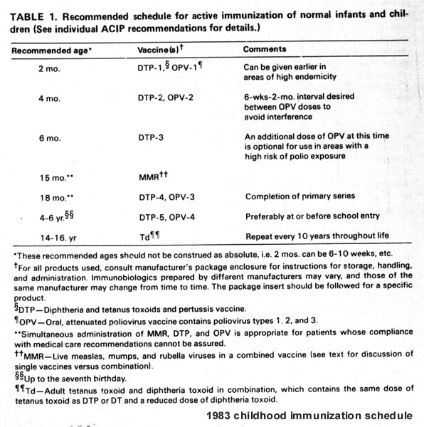
1983: A healthy-born child according to the CDC vaccination schedule [2] receives 6 vaccines in the first 15 months of life. The autism rate is 1:10,000.
2017: A healthy-born child according to the CDC vaccination schedule [3] receives 23 vaccines in the first 15 months of life. The autism rate is 1:68.
This means in the last 30 years, the prevalence of autism has risen 14,700% [3]. The projected costs for the United States would rise to more than $1 trillion by 2025 [4] if prevalence continues to rise at rates seen over last decade alone.
I want to tell you how autism comes about. Just to clarify: I am not against the concept of vaccination. I am against the toxins contained within vaccines. If you think the vaccine industry has tested all the ingredients on humans, you are deep in the woods. I invite you to examine the scientifically documented data and discover that what is happening is beyond concerning. Vaccines ARE linked to autism. And this is why.
How Vaccines Work
“Vaccines help develop immunity by imitating an infection. This type of infection, however, does not cause illness, but it does cause the immune system to produce T-lymphocytes and antibodies. Sometimes, after getting a vaccine, the imitation infection can cause minor symptoms, such as fever. Such minor symptoms are normal and should be expected as the body builds immunity. Once the imitation infection goes away, the body is left with a supply of ‘memory’ T-lymphocytes, as well as B-lymphocytes that will remember how to fight that disease in the future.” [5]
–Centers for Disease Control (CDC), CDC.GOV
A vaccine’s contents are injected into the muscle. From there it elicits a specific response from the immune system. Additives called adjuvants are put in vaccines to make the immune system response more pronounced and therefore more effective. The objective of adding adjuvants to vaccines is that adjuvants prime protective memory CD8 T-cells for future exposure. [29]. When your immune system is responding to the vaccine ingredients, it creates memory cells that will be ready to kill the real bacteria or virus when exposed to it in the future [6]. Vaccines have tiny particles of the virus or bacteria in it that your immune system recognizes as full blown real viral or bacterial threat.
Microglia – Your brain’s auto-intelligence
Figure 1 – Blood brain barrier
The brain is like a country with very tough borders. Molecules only pass through into the brain environment after proper vetting. This barrier is called the blood brain barrier. What makes this barrier effective is its tight junctions, allowing only certain molecules through [7]. This barrier separates your brain from the rest of the body, which is called the “periphery”. The reason this is key is because if you vaccinate, the ingredients of the vaccine should never even get to enter the brain.
Figure 2 – Microglia are like eyes inside your brain
If there are small foreign particles “leaking” through anyways, they get neutralized by a very effective mechanism that is controlled by a cellular structure in your brain called microglia [8] [Figure 2]. These wonder cells are more prevalent in your brain than actual brain cells. In general, microglia outnumber neuronal cells by 1.5 to 1 [9]. What makes microglia cells so interesting is that in the developing central nervous system (CNS) they can be in (pro-inflammatory) “war” mode as well as (anti-inflammatory) “beneficial” mode [10]. It makes sense that they are alerted when some foreign particles are suddenly in the brain. They recruit A LOT of other microglia to immediately help out and get rid of the danger. The “war mode” microglia cell takes care of any threats (bacterial, viral, foreign) whereas the beneficial mode actually connects neurons and is responsible for maintaining our neuronal circuits working meticulously, resulting in brain homeostasis.
Microglia cells exist in 3 states
- Resting state
- Activated pro-inflammatory state, called M1
- Beneficial anti-inflammatory / “re-constructive” state, called M2 [11]
Figure 3 – The 3 stages microglia cells
Microglia Resting State
Since microglia reside exclusively in the central nervous system, they get activated by different antigens (Figure 4, red particles). When not yet in contact with antigen, these microglia (Fig.4, green particles) are in their resting state.
Figure 4 – resting, surveillance state
What do microglia do?
Here is an example of how inflammation in the brain starts. The image below depicts a microglia (green particle) detecting a bacterial antigen called lipopolysaccharide (LPS)(red particle).
Figure 5 – Microglia (left) about to make contact with antigen LPS (right)
Lipopolysaccharide [LPS]-induced activation of microglia has been well-documented [13]. LPS is a particle from bacteria that your immune system recognizes as foreign. Once the microglia comes in contact, a powerful cry for help is the consequence, because LPS is a strong activator of microglia. The response is that the microglia secrete cytokines, little molecules that recruit other inflammatory cells to help out clean the offending antigen [25]. These will attract potent soldier like immune cells to destroy the particle. Like any war, this has a lot of innocent casualties, usually resulting in unintended neuronal inflammation.
Figure 6 – Microglia (left) releasing pro-inflammatory cytokines
LPS used to be included in vaccines, but it created so many adverse reactions (e.g. fevers), scientists re-engineered this antigen to modified versions, e.g. monophosphoryl lipid A (MPL) [13]. MPL is an active ingredient of the newborn hepatitis B vaccine and Cervarix (anti-cervical cancer vaccine). With the help of bio-synthetic engineering, more of these adjuvants that resemble LPS were developed [14]. Other microglial activators are viral particles, contained in vaccines. The MMR vaccine for example contains hemagglutinin, which has been shown to directly activate microglia [16] and is associated with autism [15].
Vaccines also carry with them even other antigens, neither viral nor bacterial. These, of course, can activate microglia as well. [14] These adjuvants are available for you to review at the CDC vaccine ingredient list [17]. Just to be clear, a vaccine is not supposed to function by intentionally activating the microglia into the pro-inflammatory M1 state. The vaccine adjuvants are NOT supposed to be in your brain at all.
Figure 8 – CDC vaccine ingredient list (cdc.gov)
Research into these adjuvants is booming, currently over 40,000 articles contain information about
“vaccine adjuvants” at the National Library of Medicine (as of 2017) [18].
In a nutshell:
Certain adjuvants in vaccines are powerful activators of brain microglia. Heavy metals like aluminum keep microglia in the activated state longer and make it difficult to switch into the anti-inflammatory state [23]. Aluminum is currently contained in DTaP, polio and Hib, Hepatitis A & B, Gardasil, Influenza and Pneumococcus [140]. As you will find out soon, there is an unreported chemical in vaccines that was found that prevents the microglia from flipping out of the inflammatory state. This chemical is called glyphosate [more below].
Microglia Beneficial M2 State
While Microglia in the M1 state are more like Pacmans eating up the offending substance and releasing pro-inflammatory cytokines, there also is a flip side to their incredible versatility. In their beneficial M2 state microglia have important physiological functions in learning and memory by promoting learning-related synapse formation. They literally connect your brain cells! This touches upon the concept of neuroplasticity [102]. The functions of the microglial M2 state are scientific and biological artistry. They elegantly orchestrate crucial steps of central nervous system development. Some of the benefits these M2 state cells provide include neuronal survival and apoptosis, axonal growth, migration of neurons, pruning of supernumerary synapses and functional maturation of developing synapses [28].
Microglia Beneficial M2 State
Similar to the M1 state, the beneficial M2 state releases anti-inflammatory cytokines and growth factors that participate in a wide range of biological responses, including increased neurogenesis and development as well as modulation of inflammation and immune responses, [104], [105], [106], [107], [108], [109]. They literally hold the keys for regulation of brain homeostasis [68].
For reference purposes, the following anti-inflammatory cytokines are released by microglia in the M2 state:
VEGF, IL-6, IL-10, PG, inducible nitric oxide synthase, IDO (immunoregulatory and proliferation-stimulating functions),IL-4, IL-10, IL-13 and TGF-beta.
Vaccine Adjuvants
In seeking to make vaccines more effective, the pharma industry’s development of vaccine adjuvants has skyrocketed. They want stronger immune responses; more specific responses; quicker responses. This is why aluminum is included in most vaccines to potentiate and prolong the overall activated M1 state [23] because the way aluminum works is by binding to the adjuvant tightly. It used to be ethylmercury (Thiomersal), but nowadays is rarely in any vaccines anymore (2017: only present in Influenza (common flu), meningococcal) [141].
The addition of an adjuvant to an existing vaccine, as has been done for influenza [19], or a switch from aluminum to a more effective adjuvant, as for hepatitis B virus (HBV) represents a substantial benefit for patients [20] [according to pharma]. There are significant numbers of people for whom current vaccines, even those using an aluminum adjuvant, do not achieve adequate immunity. Behind the curtains the pharma industry spent about 2 billion in research in 2016 to develop “better vaccines” [21].
The following are just a few adjuvants currently being added to standard vaccines [22]:
AS01 / AS02 / AS03 / AS04 / RC-529 / CpG 7909 / CpG1018 / IC31 / Imiquimod / Flagellin / AS15 / Alum / MF59 / AF03 / Virosomes / Iscomatrix / Montanide ISA51 / Montanide ISA720 / LT / LTK63
Each compound is manufactured and sold. Someone is engineering these for a reason.
AS04 for example is an approved adjuvant and is contained in the HPV and Hepatitis B vaccine and contains MPL and aluminum hydroxide.
How adjuvants push microglia into the pro-inflammatory M1 state
The following are some adjuvants where research evidence exists that they interact with microglial receptors (called Toll-Like-Receptors or TLRs). They are microglial activators and push them to be in the pro-inflammatory M1 state.
Microglia receptors that are pro-inflammatory: TLR 2 / TLR 3 / TLR 4
Figure 10a – Microglia pro-inflammatory receptors: TLR2, TLR3 and TLR4
Figure 10b: Example of the measles particle hemagluttinin getting picked up by the microglial receptor called TLR2 and converting the microglial cell into the pro-inflammatory M1 state
TLR2 receptor activators include [24]:
- Hemagluttinin (Measles) [15] (see picture above)
- Peptidoglycans (Gram+bacteria)
- Lipoproteins (variety of pathogens)
- Lipoteichoic acid (Gram+bacteria)
- Zymosan (Fungi)
- HSP70 (host, stress induced, hyperthermia, oxidative stress, and changes in pH)
- EDN (host)
TLR3 receptor activator includes:
- Double stranded rna (Rotavirus)
TLR4 receptor activators include:
- LPS (Gram-bacteria)
- Taxol (plant)
- Fusion protein (RSV)
- HSP70 (host)
- AS04 (Hepatitis B and Gardasil)
If you study this list above careful enough, you realize that if you are a perfect “vaccinator” that eventually ALL of the receptors become activated. What pushes many children into a “regression” stage is the MMR vaccine, which would be the last vaccine that docks onto the last receptor of microglia. This means the brain is in a pro-inflammatory state.
Figure 10c: Adjuvants added by year [140]
You can see that aluminum has been an ingredient for decades (Figure 10c). It just happens that these adjuvants were introduced in the mid-1990s. Interestingly enough, this is when autism rates went up (14,000%, remember? [3]). Correlation does not mean causation, but please provide a better explanation. “Better diagnostic criteria” are just not cutting it.
For example, imagine the adjuvant MPL. It is a TLR4 receptor activator. It comes along with aluminum, which binds MPL tightly and presents it to your immune system longer. So if you inject this, it’s like throwing in a piece of sugar (eg. antigen) under a bee hive, then kicking it.
Microglia take up organic mercury and convert it to the more toxic inorganic mercury [112]. Chronic methylmercury exposure leads to a large increase in activated microglia [111]. Heavy metals can therefore cause oxidative stress in neurons not only by their direct influence on sulfur metabolism but also by promoting microglia-based neuroinflammation [110]. By the way, aluminum is a heavy metal [113].
Figure 11 – Brain inflammation and autism symptoms
The response to these vaccine adjuvants is that the microglia secrete cytokines, little molecules that recruit other inflammatory cells to help out clean the offending antigen [25]. This, like any war, has a lot of innocent casualties, usually resulting in unintended neuronal activation.
In January of 2017, a Yale University study compared cytokine levels between autistic and non-autistic children and found that autistic children had statistically significantly higher levels of tumor necrosis factor alpha (TNFa) [26]. TNFa is a cytokine of microglia in the pro-inflammatory M1 state. It is known that TNF prevents conversion from the M1 (pro-inflammatory) to the M2 state (beneficial state) [27].
The brain inflammation in autism is aseptic, which means it is not caused by a true infection, but by continuously providing just enough adjuvants in vaccines to permanently keep microglia in the pro-inflammatory state. Since the microglia are not densely populating the brain stem, as they are the higher brain structures, you do not see a lot of motor disease in autism. Seizures are common, however, estimated as high as 1 in 3 [30]. In fact, you will learn how inflammation and injury in the cerebellum are likely the source of core autism symptoms (below)
Human DNA and nanoparticles in vaccines
Figure 12 – Vaccine adjuvants derived from human cells
What? Seriously? There are human cells in vaccines? The answer is YES. Even though these cells are being cultured, they still have the same source:
MRC–5 (Medical Research Council cell strain 5) is a diploid human cell culture line composed of fibroblasts derived from lung tissue of a 14 week old aborted Caucasian male fetus [31]
WI-38: The WI-38 cell line was developed in July 1962 from lung tissue taken from a therapeutically aborted fetus of about 3 months gestational age [32]
HEK-293: cells were generated in the early 1970s by transformation of cultures of normal human embryonic kidney cells with sheared adenovirus 5 DNA in Alex Van der Eb’s laboratory in Leiden, The Netherlands. The human embryonic kidney cells were obtained from previously healthy aborted fetus [33].
A recent paper has demonstrated that these human DNA particles alone are a plausible explanation to play a part in the development of autism [69]. We also know microglia sense viral RNA via its TLR3 receptor (e.g. rotavirus particles) [34].
A recent study from Italy tested vaccines and found widespread contamination by toxic aluminum salts, red blood cells of unknown origin and inorganic, foreign particle debris in aggregates, clusters and independent particulates [100]. The investigators also identified some particles embedded in a biological substrate, probably proteins, endotoxins and residues of bacteria. The researchers found contamination in 43 of the 44 vaccine samples tested. The authors stated that these contaminants should not be present in any vaccine, and that their presence was not declared by the manufacturers [101].
Apart from the inflammatory mechanisms mediated by microglia, the excitotoxin glutamate released by activated microglia is also of prime concern as excess glutamate in the brain is deleterious to neurons and synaptic connections [128].
Why is not everyone getting vaccinated become autistic?
The short answer to this question: Glyphosate
I have written several articles on how glyphosate is a key player in autism [35], [36] & [37]. Glyphosate [a.k.a Roundup, produced by Monsanto, Inc.) is the world’s most widely produced herbicide. Since 1974 in the U.S., in the form of Roundup herbicide over 1.6 billion kilograms of glyphosate have been applied, contributing to 19 % of estimated global use of glyphosate (8.6 billion kilograms) [38].
Just several months ago, Food Democracy Now tested common food products. Since any genetic modified product is by definition contaminated with glyphosate (they were genetically engineered simply to resist glyphosate, so you can spray as much on them as you like). And virtually anything non-organic has glyphosate. Soy lecithin, high fructose corn syrup, corn particles, etc. For example, cheerios were found to contain 1,125 ppb of glyphosate in them! In addition to all these tested foods found it in Bayer’s One-A-Day prenatal vitamins, newborn formula [39] and all childhood vaccines that were tested [40] (see below).
Figure 13 –Mom Across America with critical vaccine testing results
The reason this is important because of the biological consequences glyphosate has on further potentiating the above discussed mechanisms of microglial activation into the M1 state. Glyphosate was found to inhibit the P450 enzyme in the liver [41], [47]. If it inhibits P450 in the liver, it can certainly inhibit the same enzyme found in microglia in the brain, once brain access is granted. It is granted, when you don’t have someone to stop it before it gets there: the powerful compound called GcMAF. Let’s quickly learn about GcMAF, before you will see the whole picture.
What are the sources of Glyphosate in a nutshell?
GcMAF – Pacmans from your liver
The P450 enzyme is an important step in activating vitamin D3 [42] to produce a powerful compound called GcMAF. This compound eats up foreign particles in your body (like microglia in the brain). In the brain, the specific enzyme contained on microglia is called P450D6.
Figure 14 – Like microglia in the brain,GcMAF eats up foreign particles in your peripheral systems
GcMAF involves two proteins that bind it, along with the vitamin D axis, composed of the biologically active form of vitamin D (1,25(OH)(2)D3). These proteins are the vitamin D receptor (VDR) and the vitamin D binding protein that is the precursor of the vitamin D binding protein-derived macrophage activating factor, also termed GcMAF [43]. Vitamin D 25-hydroxylase is a member of the cytochrome P450 superfamily of enzymes. Found in the liver, this enzyme is a microsomal vitamin D hydroxylase that converts vitamin D into 25-hydroxyvitamin D (calcidiol), which is the major circulatory form of the vitamin [44], which will form GcMAF.
Figure 15 – How GcMAF is formed in the liver with the help of P450
GcMAF does not cause collateral damage. These little miracle molecules are like little vacuums, sucking up foreign particles that entered the system, either by injection or ingestion. They only exist in the “beneficial state.” On a molecular level these cells are probably your most powerful defense against foreign protein that leaked through the gastrointestinal tract. The mechanism to activate these GcMAF compounds utilizes Vitamin D, calcium, and your liver [again, by specifically utilizing the P450 enzyme] to make that happen.
In a nutshell:
Think of GcMAF like a monster pacman, eating up anything that is foreign, processing it, and excreting it. In theory, if you inject a child with vaccines and their livers work just fine, the GcMAF should prevent vaccine adjuvants ever reaching the brain. But what if you don’t have a lot of GcMAF because of chronic glyphosate exposure? The injected glyphosate, along with the adjuvant, are on a mission to cause trouble.
Where you could possibly be exposing your child to glyphosate
In pregnancy patients are recommended prenatal vitamins, sugar tests (corn based) and the CDC recommended flu and Tdap shots [40]. Many patients are unaware that non-organic foods are more likely than not to contain glyphosate residues [45]. The EPA has set arbitrary limits for glyphosate in our foods [71]. How about accumulative effects? Everywhere you look, glyphosate is shockingly associated with problems. Start with its production: It’s produced in the lab by fusing glycine, formaldehyde and phosphorous acid [74]. This involves an intermediate called white phosphorous [72], a highly toxic chemical used in chemical warfare [73]. Glyphosate causes bowel inflammation [46], leaky gut, exposing glyphosate to the liver, where P450 inhibiting GcMAF production will lead to a suppressed response to vaccines injected intramuscularly.
This is the reason glyphosate is now able to enter the central nervous system. Glyphosate is small enough to pass through the blood brain barrier. In order to cross the blood-brain barrier, only molecules less than 800-1000 amu (atomic mass unit) in molecular weight can get through. The molecular weight of glyphosate is about 169 amu).
Glyphosate contaminations
→ Prenatal vitamins
→ Prenatal vitamins
→ Vaccines
→ Foods
→ Newborn formulas
→ Wines, teas, sodas
→ Pregnancy glucola (results pending)?
Glyphosate inhibits the switch from pro-inflammatory M1 to anti-inflammatory M2 state
Whenever you have inflammation in the brain, your body produces a lot of receptors called CB2. This is a cannabinoid receptor. The activation of this receptor aids in neuroprotection [48], [49], [50], [51], [52], [54], [55] as also evidenced by United States Government’s Patent 6630507 [53]. It protects from glutamate damage and it has anti-oxidant activities.
The question is, where can we find this receptor in the brain? Coming full circle, the answer is: microglia.
Healthy brains don’t have CB2 receptor expression in the brain, a fact that unknown to most, yet so crucial to understand. CB2 is only is expressed when you need to “cool” the over-activation of microglia, e.g. kicking it from the pro-inflammatory M1 state into the anti-inflammatory M2 state. Normally, this happens the moment inflammation starts. The University of Michigan just recently demonstrated that in order to self-correct and cool the inflammation, the brain pulls its own endocannabinoid [anandamide] to produce a compound called 5,6-EET-EAs to activate the microglia CB2 receptor [76]. This chemical is 1000x stronger than CBD [76]. The problem is that for this conversion the enzyme P450 is required. And since glyphosate inhibits this enzyme, microglia remain permanently activated! What is mostly unknown to many is that the CB2 receptor is virtually non-existent in healthy brains [75].
“since glyphosate inhibits the P450 enzyme, microglia remain permanently activated!”
Whether the contribution of microglia actions in progressive neurodegenerative diseases is associated with an elevation in the M1 pro-inflammatory phenotype or a diminished ability of the cells to differentiate into an M2-type phenotype remains an issue under current study.
Glyphosate – It’s everywhere
23 vaccines in the first 15 months of life are on the schedule for every new member of our society. According to Stephanie Seneff, if the autism rates keep going up like they have over the last decades, we will have 1:9 children autistic by 2025 [77]. There would be too few souls to saturate our armed forces. Hence, you may conclude that autism constitutes a national security threat to the United States.
All vaccines tested had glyphosate in them [40]! You can see how a diet rich in glyphosate will put you at risk. According to CDC senior research scientist and whistleblower Dr. William Thompson [78], there was data omitted from African American children receiving the MMR vaccine. And in fact, there is an apparent link to autism.
Why would it affect African American children more so? One plausible explanation is that (according to the National Center for Children in Poverty) 33% of black children (3.6 million) live in poverty. In the 10 most populated states, rates of child poverty among black children range from 29% in California and Florida to 47% in Ohio [79]. Poverty means more processed (cheaper) food choices, which means more GMO derived foods, which means more glyphosate.
VAXXED is an excellent documentary that demonstrates the fraud and cover up [80]. Del Bigtree, Andrew Wakefield and Polly Tommey are travelling the world to raise awareness in their crusade for truth, risking so much for the sake of our children.
The accumulative amounts of glyphosate contained within these vaccines that big pharma regularly pushes onto newborns are concerning. Just hours after birth, vaccination with Hepatitis B is on the agenda. As discussed above, it contains AS04 (activator) in addition to glyphosate (a beneficial state inhibitor) [24]. Furthermore, breast milk and formula may contain glyphosate [81]&[82]. Hepatitis B and Rotavirus activate 2 of the 3 microglial TLR receptors, the MMR particle activates the third [24].
It is important to note that because glyphosate is never used alone in industry, the detection of glyphosate may be an indicator of the presence of many other co-formulants in glyphosate-based herbicides which have recently been shown by French scientist Seralini’s team to be endocrine disruptors and up to 1000 times more toxic than glyphosate alone [67].
The Cerebellum – Microglia Central Command
The cerebellum is involved in a substantial number of complex functions, ranging from the coordination of movements to language processing, spatial cognition, and other higher cognitive and affective functions [114], [115], [116]. It undergoes major growth and synaptic reorganization after birth, leading to the development of cerebellar circuits which are involved in motor and cognitive functions [130]. Damage to the cerebellum typically leads to motor deficits, but it can also result in cognitive impairments such as loss of working memory and verbal fluency, and they are associated conditions such as autism and dyslexia.
Gray matter in the cerebella of subjects with autism
Microglial activation has been documented in the cerebella of subjects by autism [119], [120], [121], [122], [126]. Multiple studies have demonstrated significant reductions in gray matter in the cerebella of subjects with autism [123], [124], [125]. These reductions correlated with scores assessing repetitive and stereotyped behavior, and social behavior and communication [124]. Autopsy studies performed on autistic brains revealed marked activation of microglia [127] and sustained neurological inflammatory responses due to microglial activation in cortical and subcortical white matter as well as in the cerebellum [119]. More evidence that the cerebellum is likely the target of vaccine adjuvants is that the lateral hemispheres engaged in cognitive processing mature particularly late [129]. This renders the cerebellum vulnerable to environmental influences such as vaccine adjuvants. In conclusion, the cerebellum is the site of extensive pathology in autism spectrum disorders including abnormalities in cerebellar structural brain connections to other brain sites as well as in various proteins and neurotransmitters affecting multiple functional domains [131-139].
Cannabis – Microglia M2 activator
So far we have learned that the vaccine adjuvants (aluminum + antigen) activate microglia to be in a pro-inflammatory M1 state. The addition of glyphosate inhibits our own endocannabinoids to reverse this process. The result is irreversible activation of microglia, leaving the brain in a permanent inflammatory state. The result is autism, where memory formation is greatly impaired and immune dysregulation is the consequence. Bowel problems, parasite overgrowths and metabolic malfunctions are often evident in children affected with autism. These bacterial and parasitic overgrowths in the body provide possibly even more cytokines to the brain, keeping the microglia in their activated M1 state.
So why is cannabis so interesting? I have extensively covered phytocannabinoid mechanisms here:
Endocannabinoid System Autism & Cannabis Part 1 [95]
Phytocannabinoids Role in Autism Spectrum Disorder Therapies Part 2 [96] Practical Approach to Cannabis Based ASD Therapies Part 3 [97] The Autism Brain – How glyphosate destroys “super-cannabinoid” production [98] What glyphosate does to your brain [99]
How exactly does cannabis interact with microglia? The answer is simple, they have receptors for it, namely CB2 and TRVP. THC has been shown to reduce the amount of pro-inflammatory cytokines via CB2 interaction [52]. In fact, CB2 receptors have rarely been observed in neurons and are expressed primarily in microglial cells [117], [118].
The tremendous effects of phyto (plant) cannabinoids on microglia were recently reported by a study from the University of Madrid in Spain [57].
Evidence of microglial control by cannabis
- CB2 receptor is upregulated once microglia undergo transformation into the pro-inflammatory cell [56]. This means that the brain is self-regulating. Remember, glyphosate, contained in our diets (and every vaccine) inhibits this process.
- CB2 activation via anandamide suppresses pro-inflammatory cytokines, TNF-alpha and nitrous oxide [58]
- Cannabinoids potentiate the production of anti-inflammatory cytokine IL-6 [60]
- Cannabinoids prevents pro-inflammatory cytokine production which was induced by LPS [61]
- Endo-and phytocannabinoids activate CB2 to induce cell migration (to tell other microglia to help with the clean-up and restructuring) [62][63],[64]
- Cannabinoid activates CB2 on microglia to increase beneficiary M2 state proliferation [65]
- THC-mediated CB2 activation resulting in fewer number of microglial cells and fewer number of degenerating neurons [66]
Cannabis remains a Schedule-I drug (the most harmful of all, according to the DEA), and therefore clinical trials are impossible to conduct. This is why it is imperative the federal government de-schedules cannabis.
Resources
Great resources about cannabis in autism:
- MAMMAs (Mothers Advocating Medical Marijuana for Autism) (Facebook [87] and website)
- Mieko Hester Perez, The Unconventional Foundation for Autism [93]
- Whole Plant Access for Autism (Facebook [86])
Great resources about glyphosate:
- March against Monsanto [89] & [90]
- Moms across America [91] & [92]
- Stephanie Seneff, MIT research [77]
National Library of Medicine Toxnet Database [94]
Suggested future implications
- Ban genetic modification of food and fund organic farming (join the March against Monsanto movement) [84]
- Avoid GMO products and buy organic
- Demand “clean” vaccines [85]
- Demand a vaccine schedule that makes scientific sense (e.g. there is no good reason to vaccinate a newborn with Hepatitis B when both mother and father are not affected with hepatitis B)
- Supoena Dr. William Thompson from the CDC and analyze omitted data to have a dialogue with the strong pro-vaxx movement to reformulate the ingredients
- De-schedule cannabis just because there is absolutely no reason to schedule it in the first place. We have overwhelming scientific evidence of it being therapeutic in various disease processes
This article will be heavily criticized. Most likely it has flaws and errors, but overall I have referenced over 140 sources supporting the above picture, drawing the core principles of autism pathophysiology. Please read them in their entirety before debunking this information.
I claim that withholding cannabis as a treatment modality is a human rights violation, as there is no good evidence that it can cause harm. According to the World Health Organization, almost six million people die from tobacco use and 2.5 million from harmful use of alcohol each year worldwide, yet, both are legal. Most importantly, however, is that we are causing harm to our children with reckless vaccination schedules that are poorly studied and tested on humans. This needs to stop. Demand change. Today.
~ Dr. Christian Bogner, MD
A special thanks to the research community and activists who are all striving to put an end to this epidemic:
Chief investigator Joe Stone, Dwight Zahringer, Dr. Jeff Bradstreet, Thom and Candice Bradstreet, Dr. Stephanie Seneff, Dr. Lester Grinspoon, Michael Komorn, Tami Canal (March against Monsanto), Zen Honeycutt (Moms across America), Sterling Hill, Kerry Rivera, Jason Cranford, Richard Haines, MAMMA USA, Mieko Hester Perez (uf4a.org),Andrew Wakefield, Del Bigtree, Polly Tommey, Leah Hochbaum, Abigail Dar, Terri and Ed Arranga and all the parents and children standing with us. Thank you Kristin Thomas for proofreading.
References
[6] Coffman RL, Sher A, Seder RA. Vaccine adjuvants: putting innate immunity to work. Immunity. 2010;33(4):492-503.
[7] The Blood-Brain Barrier in Health and Disease, Volume One: Morphology, Biology and Immune Function, CRC Press (June 23, 2015)
[8] Ransohoff RM, El khoury J. Microglia in Health and Disease. Cold Spring Harb Perspect Biol. 2015;8(1):a020560.
[9] Azevedo FA, Carvalho LR, Grinberg LT, et al. Equal numbers of neuronal and nonneuronal cells make the human brain an isometrically scaled-up primate brain. J Comp Neurol. 2009;513(5):532-41.
[10] Glial Physiology and Pathophysiology, Wiley-Blackwell; 1 edition (April 15, 2013)
[11] Cherry JD, Olschowka JA, O’banion MK. Neuroinflammation and M2 microglia: the good, the bad, and the inflamed. J Neuroinflammation. 2014;11:98.
[12]Ransohoff RM, El khoury J. Microglia in Health and Disease. Cold Spring Harb Perspect Biol. 2015;8(1):a020560
[13]Zariri A, Pupo E, Van riet E, Van putten JP, Van der ley P. Modulating endotoxin activity by combinatorial bioengineering of meningococcal lipopolysaccharide. Sci Rep. 2016;6:36575.
[14] Coffman RL, Sher A, Seder RA. Vaccine adjuvants: putting innate immunity to work. Immunity. 2010;33(4):492-503.
[15] Singh VK, Lin SX, Newell E, Nelson C. Abnormal measles-mumps-rubella antibodies and CNS autoimmunity in children with autism. J Biomed Sci. 2002;9(4):359-64.
[16] Bieback K, Lien E, Klagge IM, et al. Hemagglutinin protein of wild-type measles virus activates toll-like receptor 2 signaling. J Virol. 2002;76(17):8729-36.
[19] Podda A. The adjuvanted influenza vaccines with novel adjuvants: experience with the MF59-adjuvanted vaccine. Vaccine. 2001;19(17-19):2673-80.
[20] Beran J. Safety and immunogenicity of a new hepatitis B vaccine for the protection of patients with renal insufficiency including pre-haemodialysis and haemodialysis patients. Expert Opin Biol Ther. 2008;8(2):235-47.
[22] De gregorio E, Caproni E, Ulmer JB. Vaccine adjuvants: mode of action. Front Immunol. 2013;4:214.
[23] Exley C, Siesjö P, Eriksson H. The immunobiology of aluminium adjuvants: how do they really work?. Trends Immunol. 2010;31(3):103-9.
[24] Cancer and Inflammation Mechanisms: Chemical, Biological, and Clinical Aspects 1st Edition (March 31, 2014)
[25] Gertig U, Hanisch UK. Microglial diversity by responses and responders. Front Cell Neurosci. 2014;8:101.
[26] Guloksuz SA, Abali O, Aktas cetin E, et al. Elevated plasma concentrations of S100 calcium-binding protein B and tumor necrosis factor alpha in children with autism spectrum disorders. Rev Bras Psiquiatr. 2017;
[27] Kroner A, Greenhalgh AD, Zarruk JG, Passos dos santos R, Gaestel M, David S. TNF and increased intracellular iron alter macrophage polarization to a detrimental M1 phenotype in the injured spinal cord. Neuron. 2014;83(5):1098-116.
[28] Mosser CA, Baptista S, Arnoux I, Audinat E. Microglia in CNS development: Shaping the brain for the future. Prog Neurobiol. 2017;
[29] Macleod MK, Mckee AS, David A, et al. Vaccine adjuvants aluminum and monophosphoryl lipid A provide distinct signals to generate protective cytotoxic memory CD8 T cells. Proc Natl Acad Sci USA. 2011;108(19):7914-9.
[30] Tuchman R. Autism and epilepsy: what has regression got to do with it?. Epilepsy Curr. 2006;6(4):107-11.
[34] Ge Y, Mansell A, Ussher JE, et al. Rotavirus NSP4 Triggers Secretion of Proinflammatory Cytokines from Macrophages via Toll-Like Receptor 2. J Virol. 2013;87(20):11160-7.
[38] Benbrook CM. Trends in glyphosate herbicide use in the United States and globally. Environ Sci Eur. 2016;28(1):3.
[39] Samsel A, Seneff S. Glyphosate, pathways to modern diseases III: Manganese, neurological diseases, and associated pathologies. Surg Neurol Int. 2015;6:45.
[41] Samsel A, Seneff S. Glyphosate’s Suppression of Cytochrome P450 Enzymes and Amino Acid Biosynthesis by the Gut Microbiome: Pathways to Modern Diseases. Entropy 2013, 15(4), 1416-1463
[42] Christakos S, Dhawan P, Verstuyf A, Verlinden L, Carmeliet G. Vitamin D: Metabolism, Molecular Mechanism of Action, and Pleiotropic Effects. Physiol Rev. 2016;96(1):365-408.
[43] Thyer L, Ward E, Smith R, et al. A novel role for a major component of the vitamin D axis: vitamin D binding protein-derived macrophage activating factor induces human breast cancer cell apoptosis through stimulation of macrophages. Nutrients. 2013;5(7):2577-89.
[46] National Toxicology Data Network, Glyphosate. https://toxnet.nlm.nih.gov/cgi-bin/sis/search/a?dbs+hsdb:@term+@DOCNO+3432
[47] World Health Organization WHO/ International Programme on Chemical Safety; Environmental Health Criteria 159, Glyphosate, (1994)
[48] Cassano T, Calcagnini S, Pace L, De marco F, Romano A, Gaetani S. Cannabinoid Receptor 2 Signaling in Neurodegenerative Disorders: From Pathogenesis to a Promising Therapeutic Target. Front Neurosci. 2017;11:30.
[49] Fernández-trapero M, Espejo-porras F, Rodríguez-cueto C, et al. Up-regulation of CB2 receptors in reactive astrocytes in canine degenerative myelopathy, a disease model of amyotrophic lateral sclerosis. Dis Model Mech. 2017;
[50] Navarro G, Morales P, Rodríguez-cueto C, Fernández-ruiz J, Jagerovic N, Franco R. Targeting Cannabinoid CB2 Receptors in the Central Nervous System. Medicinal Chemistry Approaches with Focus on Neurodegenerative Disorders. Front Neurosci. 2016;10:406.
[51] Javed H, Azimullah S, Haque ME, Ojha SK. Cannabinoid Type 2 (CB2) Receptors Activation Protects against Oxidative Stress and Neuroinflammation Associated Dopaminergic Neurodegeneration in Rotenone Model of Parkinson’s Disease. Front Neurosci. 2016;10:321.
[52] Xie J, Xiao D, Xu Y, et al. Up-regulation of immunomodulatory effects of mouse bone-marrow derived mesenchymal stem cells by tetrahydrocannabinol pre-treatment involving cannabinoid receptor CB2. Oncotarget. 2016;7(6):6436-47.
[53] US patent 6630507,Cannabinoids as antioxidants and neuroprotectants, https://docs.google.com/viewer?url=patentimages.storage.googleapis.com/pdfs/US6630507.pdf
[54] Fernández-Ruiz JJ, Gonzalez S, Romero J, Ramos JA. Cannabinoids in neurodegeneration and neuroprotection Cannabinoids as Therapeutics 2005,Birkhäuser Verlag: Switzerland; 79–109.109In: Mechoulam R (ed)
[55] Walsh SK, Hepburn CY, Keown O, et al. Pharmacological profiling of the hemodynamic effects of cannabinoid ligands: a combined in vitro and in vivo approach. Pharmacol Res Perspect. 2015;3(3):e00143.
[56] Carlisle SJ, Marciano-Cabral F, Staab A, Ludwick C, Cabral GA (2002). Differential expression of the CB2 cannabinoid receptor by rodent macrophages and macrophage-like cells in relation to cell activation. Int Immunopharmacol 2: 69–82
[57] Benito C, Tolón RM, Pazos MR, Núñez E, Castillo AI, Romero J. Cannabinoid CB2 receptors in human brain inflammation. Br J Pharmacol. 2008;153(2):277-85.
[58] Molina-Holgado F, Lledo A, Guaza C. Anandamide suppresses nitric oxide and TNF-alpha responses to Theiler’s virus or endotoxin in astrocytes. Neuroreport. 1997;8:1929–1933.
[59] Walter L, Franklin A,Witting A,Wade C, Xie Y, Kunos G et al. (2003).Nonpsychotropic cannabinoid receptors regulate microglial cell migration. J Neurosci 23: 1398–1405.
[60] Molina-Holgado F, Molina-Holgado E, Guaza C. The endogenous cannabinoid anandamide potentiates interleukin-6 production by astrocytes infected with Theiler’s murine encephalomyelitis virus by a receptor-mediated pathway. FEBS Lett. 1998;433:139–142.
[61] Puffenbarger RA, Boothe AC, Cabral GA. Cannabinoids inhibit LPS-inducible cytokine mRNA expression in rat microglial cells. Glia. 2000;29:58–69.
[62] Walter L, Franklin A,Witting A,Wade C, Xie Y, Kunos G et al. (2003). Nonpsychotropic cannabinoid receptors regulate microglial cell migration. J Neurosci 23: 1398–1405.
[63] Klein TW, Lane B, Newton CA, Friedman H. The cannabinoid system and cytokine network. Proc Soc Exp Biol Med. 2000;225:1–8.
[64] Franklin A, Stella N (2003). Arachidonylcyclopropylamide increases microglial cell migration through cannabinoid CB2 and abnormal cannabidiol-sensitive receptors. Eur J Pharmacol 474: 195–198.
[65] Carrier EJ, Kearn CS, Barkmeier AJ, Breese NM, YangW, Nithipatikom,K et al. (2004). Cultured rat microglial cells synthesize the endocannabinoid 2-arachidonylglycerol, which increases proliferation via a CB2 receptor-dependent mechanism. Mol Pharmacol 65: 999–1007.
[66] Kreutz S, Koch M, Ghadban C, Korf HW, Dehghani F (2007). Cannabinoids and neuronal damage: differential effects of THC,AEA and 2-AG on activated microglial cells and degenerating neurons in excitotoxically lesioned rat organotypic hippocampal slice cultures. Exp Neurol 203: 246–257.
[68] Glial Physiology and Pathophysiology, Wiley-Blackwell; 1 edition (April 15, 2013
[75] Onaivi ES, Ishiguro H, Gu S, Liu QR. CNS effects of CB2 cannabinoid receptors: beyond neuro-immuno-cannabinoid activity. J Psychopharmacol (Oxford). 2012;26(1):92-103.
[76] Snider NT, Sikora MJ, Sridar C, Feuerstein TJ, Rae JM, Hollenberg PF. The endocannabinoid anandamide is a substrate for the human polymorphic cytochrome P450 2D6. J Pharmacol Exp Ther. 2008;327(2):538-45.
[82] Samsel A, Seneff S. Glyphosate, pathways to modern diseases III: Manganese, neurological diseases, and associated pathologies. Surg Neurol Int. 2015;6:45.
[103] Handbook of the Cerebellum and Cerebellar Disorders, Manto, 2013 [Springer]
[104] Aarum J, Sandberg K, Haeberlein SLB, Persson MAA (2003) Migration and differentiation of neural precursor cells can be directed by microglia. Proc Natl Acad Sci USA 100:15983–15988
[105] Morgan SC, Taylor DL, Pocock JM (2004) Microglia release activators of neuronal proliferation mediated by activation of mitogen-activated protein kinase, phosphatidylinositol-3-kinase/Akt and delta–Notch signalling cascades. J Neurochem 90:89–101
[106] Battista D, Ferrari CC, Gage FH, Pitossi FJ (2006) Neurogenic niche modulation by activated microglia: transforming growth factor β increases neurogenesis in the adult dentate gyrus. Eur J Neurosci 23:83–93
[107] Butovsky O, Ziv Y, Schwartz A, Landa G, Talpalar AE, Pluchino S et al (2006) Microglia activated by IL-4 or IFN-γ differentially induce neurogenesis and oligodendrogenesis from adult stem/ progenitor cells. Mol Cell Neurosci 31:149–160
[108] Ziv Y, Ron N, Butovsky O, Landa G, Sudai E, Greenberg N et al (2006) Immune cells contribute to the maintenance of neurogenesis and spatial learning abilities in adulthood. Nat Neurosci 9:268–275
[109] Vukovic J, Colditz MJ, Blackmore DG, Ruitenberg MJ, Bartlett PF (2012) Microglia modulate hippocampal neural precursor activity in response to exercise and aging. J Neurosci 32: 6435–6443
[110] The Neurochemical Basis of Autism, Springer; 2010 edition (March 10, 2010)
[111] Charleston JS, Bolender RP, Mottet NK, Body RL, Vahter ME, Burbacher TM. (1994) Increases in the number of reactive glia in the visual cortex of Macaca fascicularis following subclinical long-term methyl mercury exposure. Toxicol Appl Pharmacol 129:196–206.
[112] Charleston JS, Body RL, Mottet NK, Vahter ME, Burbacher TM. (1995) Autometallographic determination of inorganic mercury distribution in the cortex of the calcarine sulcus of the monkey Macaca fascicularis following long-term subclinical exposure to ethylmercury and mercuric chloride. Toxicol Appl Pharmacol 132:325–333.
[114] Baudouin, S. J., Gaudias, J., Gerharz, S., Hatstatt, L., Zhou, K., Punnakkal, P., et al. (2012). Shared synaptic pathophysiology in syndromic and nonsyndromic rodent models of autism. Science, 338(6103), 128–132.
[115] Schmahmann, J. D. (2010). The role of the cerebellum in cognition and emotion: personal reflections since 1982 on the dysmetria of thought hypothesis, and its historical evolution from theory to therapy. Neuropsychology review, 20(3), 236–260.
[116] Tsai, P. T., Hull, C., Chu, Y., Greene-Colozzi, E., Sadowski, A. R., Leech, J. M., et al. (2012). Autistic-like behaviour and cerebellar dysfunction in Purkinje cell Tsc1 mutant mice. Nature, 488(7413), 647–651.
[117] Carrier EJ, Kearn CS, Barkmeier AJ, Breese NM, Yang W, Nithipatikom K, Pfister SL, Campbell WB, Hillard CJ (2004) Cultured rat microglial cells synthesize the endocannabinoid 2-arachidonylglycerol, which increases proliferation via a CB2 receptor-dependent mechanism. Mol Pharmacol 65:999–1007
[118] Gong JP, Onaivi ES, Ishiguro H, Liu QR, Tagliaferro PA, Brusco A, Uhl GR (2006) Cannabinoid CB2 receptors: immunohistochemical localization in rat brain. Brain Res 1071:10–23
[119] Vargas DL, Nascimbene C, Krishnan C et al (2005) Neuroglial activation and neuroinflammation in the brain of patients with autism. Ann Neurol 57:67–81
[120] Chaste P, Leboyer M: Autism risk factors: Genes, environment, and gene-environment interactions. Dialogues Clin Neurosci 14:281–292, 2012.
[121] Jyonouchi H, Sun S, Le H: Proinflammatory and regulatory cytokine production associated with innate and adaptive immune responses in children with autism spectrum disorders and developmental regression.J Neuroimmunol 120:170–179, 2001.
[122] Ashwood P, Krakowiak P, Hertz-Picciotto I, Hansen R, Pessah I, Van de Water J: Elevated plasma cytokines in autism spectrum disorders provide evidence of immune dysfunction and are associated with impaired behavioral outcome. Brain Behav Immun 25:40–45, 2011.
[123] McAlonan GM, Cheung V, Cheung C et al (2005) Mapping the brain in autism: a voxel-based MRI study of volumetric differences and intercorrelations in autism. Brain 128:268–276
[124] Rojas DC, Peterson E, Winterrowd E et al (2006) Regional gray matter volumetric changes in autism associated with social and repetitive behavior symptoms. BMC Psychiatry 6:56
[125] Toal F, Bloemen OJ, Deeley Q et al (2009) Psychosis and autism: magnetic resonance imaging study of brain anatomy. Br J Psychiatry 194:418–425
[126] Morgan JT, Chana G, Pardo CA, Achim C, Semendeferi K, Buckwalter J, Courchesne E,Everall IP. Microglial activation and increased microglial density observed in the dorsolateral prefrontal cortex in autism. Biol. Psychiatry (2010) 68:368-376.
[127] Pardo CA, Vargas DL, Zimmerman AW. Immunity, neuroglia and neuroinflammation in autism. Int. Rev. Psychiatry (2005) 17:485-495.
[128] Blaylock RL. Chronic Microglial Activation and Excitotoxicity Secondary to Excessive Immune Stimulation: Possible Factors in Gulf War Syndrome and Autism. J. Am. Phys.Surg. (2004) 9:46-51.
[129] Tiemeier H, Lenroot RK, Greenstein DK et al (2010) Cerebellum development during childhood and adolescence: a longitudinal morphometric MRI study. Neuroimage 49(1):63–70
[130] Perez-pouchoulen M, Vanryzin JW, Mccarthy MM. Morphological and Phagocytic Profile of microglia in the Developing Rat Cerebellum(1,2,3). eNeuro. 2015;2(4)
[131] Fatemi SH, Stary JM, Halt AR, Realmuto G (2001) Dysregulation of Reelin and Bcl-2 proteins in autistic cerebellum. J Autism Dev Disord 6:529–535
[132] Fatemi SH, Halt AR, Realmuto G, Earle J, Kist DA, Thuras P, Merz A (2002a) Purkinje cell size is reduced in cerebellum of patients with autism. Cell Mol Neurobiol 22:171–175
[133] Fatemi SH, Halt AR, Stary JM, Kanodia R, Schulz SC, Realmuto GR (2002b) Glutamic acid decarboxylase 65 and 67 kDa proteins are reduced in autistic parietal and cerebellar corticies. Biol Psychiatry 52:805–810
[134] Fatemi SH, Snow AV, Stary JM, Araghi-Niknam M, Reutiman TJ, Lee S, Brooks AI, Pearce DA (2005) Reelin signaling is impaired in autism. Biol Psychiatry 57:777–787
[135] Fatemi SH, Reutiman TJ, Folsom TD, Thuras PD (2009a) GABA(A) receptor downregulation in brains of subjects with autism. J Autism Dev Disord 39:223–230
[136] Fatemi SH, Folsom TD, Reutiman TJ, Thuras PD (2009b) Expression of GABA(B) receptors is altered in brains of Subjects with autism. Cerebellum 8:64–69
[137] Fatemi SH, Reutiman TJ, Folsom TD, Rooney RJ, Patel DH, Thuras PD (2010) mRNA and protein levels for GABA(A) alpha 4, alpha 5, beta 1, and GABA(B)R1 receptors are altered in brains from subjects with autism. J Autism Dev Disord 40:743–750
[138] Fatemi SH, Folsom TD, Kneeland RE, Liesch SB (2011) Metabotropic glutamate receptor 5 upregulation in children with autism is associated with underexpression of both Fragile X mental retardation protein and GABAA receptor beta 3 in adults with autism. Anat Rec 294:1635–1645
[139] Fatemi SH, Aldinger KA, Ashwood P, Bauman ML, Blaha CD, Blatt GJ, Chauhan A, Chauhan V, Dager SR, Dickson PE, Estes AM, Goldowitz D, Heck DH, Kemper TL, King BH, Martin LA, Millen KJ, Mittleman G, Mosconi MW, Persico AM, Sweeney JA, Webb SJ, Welsh JP (2012) Consensus paper: pathological role of the cerebellum in autism. Cerebellum 11:777–807
[140] Available at: http://www.sciencedirect.com/science/article/pii/S2210762211000052. Accessed March 14, 2017.
1 comment:
QUANTUM BINARY SIGNALS
Get professional trading signals delivered to your mobile phone every day.
Follow our trades right now & gain up to 270% daily.
Post a Comment